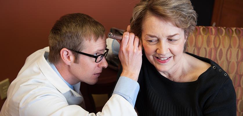
Video Otoscopy
We'll examine your ears to check for excessive wax and other problems.
Otoscopy or Video Otoscopy
Video otoscopy is a diagnostic technique that allows the visualization of the ear canal and the tympanic membrane using a small camera attached to a handheld device. It is a useful tool for assessing the health and function of the ear, as well as for detecting and treating various ear conditions.
Video otoscopy has several advantages over conventional otoscopy, such as:
- It provides a magnified and high-resolution image of the ear structures, which can reveal subtle abnormalities that might be missed by the naked eye.
- It allows the recording and storage of the images for documentation and comparison purposes, which can facilitate the diagnosis and follow-up of ear problems.
- It enables the sharing and consultation of the images with other professionals or specialists, which can improve the quality and accuracy of the care.
- It enhances the communication and education of the patients, who can see their own ear anatomy and pathology on a screen and better understand their condition and treatment options.
Video otoscopy can be performed in various settings, such as primary care clinics, audiology centers, ENT offices, or even at home with portable devices. It is a safe and painless procedure that usually takes only a few minutes. The video otoscope is gently inserted into the ear canal and moved around to examine different areas of the ear. The images are displayed on a monitor or a smartphone screen in real time.
Video otoscopy can help diagnose and treat a wide range of ear conditions, such as:
- Earwax impaction: Video otoscopy can help identify and remove excessive or impacted earwax, which can cause hearing loss, discomfort, or infection.
- Otitis externa: Video otoscopy can help detect and treat inflammation or infection of the outer ear canal, which can cause pain, itching, discharge, or swelling.
- Otitis media: Video otoscopy can help identify and manage inflammation or infection of the middle ear, which can cause fever, hearing loss, fluid accumulation, or perforation of the tympanic membrane.
- Tympanic membrane perforation: Video otoscopy can help assess and monitor the size and location of a hole in the tympanic membrane, which can result from trauma, infection, or pressure changes.
- Tympanosclerosis: Video otoscopy can help detect and evaluate the presence of calcium deposits on the tympanic membrane or the middle ear bones, which can cause hearing loss or stiffness of the ossicles.
- Cholesteatoma: Video otoscopy can help identify and treat a benign growth of skin cells in the middle ear or mastoid bone, which can cause hearing loss, discharge, infection, or damage to the facial nerve.
- Otosclerosis: Video otoscopy can help diagnose and monitor a condition that causes abnormal bone growth in the middle ear, which can cause hearing loss or tinnitus.
- Eustachian tube dysfunction: Video otoscopy can help evaluate and manage a condition that affects the function of the tube that connects the middle ear to the throat, which can cause pressure changes, fluid accumulation, or infection in the middle ear.
Video otoscopy is a valuable technique that can improve the diagnosis and treatment of various ear conditions. It is also a powerful tool for patient education and empowerment. By using video otoscopy, both clinicians and patients can benefit from a better understanding and management of ear health.
Your audiologist will examine your ears with an otoscope to check for excessive wax and other problems. If we find something of concern, we may use a video otoscope which is connected to a TV or computer screen, to show you the affected area. Of course we will recommend you see an otolaryngologist (physician that specializes in matters of the ears, nose, and throat) if warranted.
Schedule your consultation at our Huntington office or to find out more about hearing tests, contact us at
(631) 271-6263.